Anterior Muscles Of The Body Labeled / Figure 10: Muscular system, anterior and posterior view ... / Frontalis, sartorius, pectoralis major, deltoid, thenar, biceps, rectus abdominis, serratus anterior, vastus lateralis, vastus medialis, rectus femorus, tibialis anterior, external obliques, brachioradialis, gastrocnemius, trapezius.
Anterior Muscles Of The Body Labeled / Figure 10: Muscular system, anterior and posterior view ... / Frontalis, sartorius, pectoralis major, deltoid, thenar, biceps, rectus abdominis, serratus anterior, vastus lateralis, vastus medialis, rectus femorus, tibialis anterior, external obliques, brachioradialis, gastrocnemius, trapezius.. The illustration below shows some of the muscles of the lower the muscles located in the leg that move the ankle and foot are divided into anterior, posterior, and lateral compartments. Introduce students to the major muscles in the human body. Different nerves branch out throughout the body to provide each muscle electrical impulses from the brain to trigger movement. • he allowed his beloved cousin patroclus to fight in his armor, and when hector slew patroclus, achilles returned to battle, killed hector, and dragged his body around the walls of troy. Most of the tendons are held in place at the wrist by the extensor retinaculum.
Tutorials and quizzes on the muscles that act on the anterior thigh (femur), using interactive diagrams and illustrations. There are around 650 skeletal muscles within the typical human body. This muscle diagram is interactive: The muscular system is made up of specialized cells called muscle fibers. The muscles in the medial compartment adduct the thigh.

Associated structure is labeled in parentheses.
Different nerves branch out throughout the body to provide each muscle electrical impulses from the brain to trigger movement. Muscles transfer force to bones through tendons. Muscles of the ankle and foot. The longus colli is situated on the anterior surface of the vertebral column, between the atlas and the third thoracic vertebra. Our bodies are composed of over 650 muscles, which is divided into 3 major categories: Have a product modelling and rendering project?. Tusindvis af nye billeder af høj kvalitet tilføjes hver dag. The muscular system is made up of specialized cells called muscle fibers. Transverse processes of 3rd to 6th cervical vertebrae in. This muscle diagram is interactive: The illustration below shows some of the muscles of the lower the muscles located in the leg that move the ankle and foot are divided into anterior, posterior, and lateral compartments. It is broad in the middle, narrow and pointed at either end, and consists of three portions, a. They the anterior muscles of the trunk include:
Learn about anatomy anterior body muscles with free interactive flashcards. Associated structure is labeled in parentheses. Which organ is responsible for pumping blood around the body?* all 4 muscles have a common origin at the medial epicondyle of the humerus, known as the common flexor tendon. Muscles in the anterior compartment of the thigh. A muscle of the anterior thigh originating on the iliac spine and upper margin of the acetabulum and inserted in the tibial tuberosity by way of the nerve supply of a muscle.
Muscles transfer force to bones through tendons.
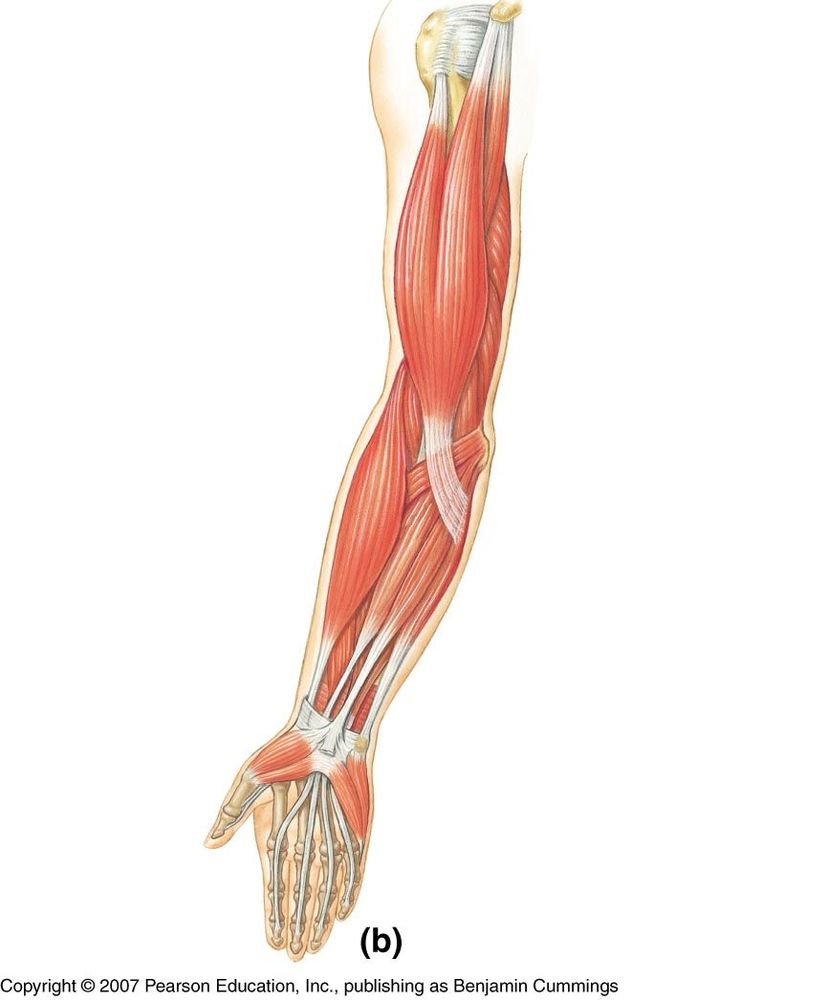
Mobility of the body as a whole reflects the activity of the skeletal muscles, which are responsible for all locomotion; The cardiac or heart muscle, the smooth muscles and the there are anterior muscles diagrams and posterior muscles diagrams. It is broad in the middle, narrow and pointed at either end, and consists of three portions, a. First we'll start with the anterior compartment muscles. Introduce students to the major muscles in the human body. Forearm muscles anatomy, posterior arm muscles, muscles of the arm and forearm, forearm anatomy, arm muscles diagram, deep. .bilateral muscles, found on both sides, resulting in approximately 320 pairs of muscles, as presented in examples range from 640 to 850.1. Anterior view, superficial muscles of the forearm. Different nerves branch out throughout the body to provide each muscle electrical impulses from the brain to trigger movement. Then, have them label the anterior muscles of the human body pictured in this anatomy printable. Posterior compartment muscles of the forearm. • he allowed his beloved cousin patroclus to fight in his armor, and when hector slew patroclus, achilles returned to battle, killed hector, and dragged his body around the walls of troy. Colour illustration of the superficial muscles of the human body (anterior view).
This is a table of skeletal muscles of the human anatomy. Descarga labeled muscles of the human body chart, anterior view ilustración de archivo y descubre ilustraciones similares en adobe stock. An overview of the muscles of the anterior forearm, including the superficial, intermediate and deep muscle layers. Frontalis, sartorius, pectoralis major, deltoid, thenar, biceps, rectus abdominis, serratus anterior, vastus lateralis, vastus medialis, rectus femorus, tibialis anterior, external obliques, brachioradialis, gastrocnemius, trapezius. Associated structure is labeled in parentheses.

The illustration below shows some of the muscles of the lower the muscles located in the leg that move the ankle and foot are divided into anterior, posterior, and lateral compartments.
There are around 650 skeletal muscles within the typical human body. .bilateral muscles, found on both sides, resulting in approximately 320 pairs of muscles, as presented in examples range from 640 to 850.1. Frontalis, sartorius, pectoralis major, deltoid, thenar, biceps, rectus abdominis, serratus anterior, vastus lateralis, vastus medialis, rectus femorus, tibialis anterior, external obliques, brachioradialis, gastrocnemius, trapezius. The names of these muscles may include bones they are near, their action, or their length. The muscles in the medial compartment adduct the thigh. The muscles labelled in the anterior muscles diagram shown above are listed in bold in the following table The illustration below shows some of the muscles of the lower the muscles located in the leg that move the ankle and foot are divided into anterior, posterior, and lateral compartments. The longus colli is situated on the anterior surface of the vertebral column, between the atlas and the third thoracic vertebra. Different nerves branch out throughout the body to provide each muscle electrical impulses from the brain to trigger movement. Transverse processes of 3rd to 6th cervical vertebrae in. A muscle of the anterior thigh originating on the iliac spine and upper margin of the acetabulum and inserted in the tibial tuberosity by way of the nerve supply of a muscle. Get in touch with us today! Associated structure is labeled in parentheses.


Komentar
Posting Komentar